< img height="1" width="1" style="display:none" src="https://www.facebook.com/tr?id=788420016578267&ev=PageView&noscript=1" />

Macular Disease

1. What is Macular Degeneration

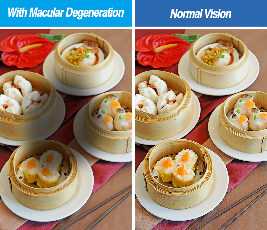
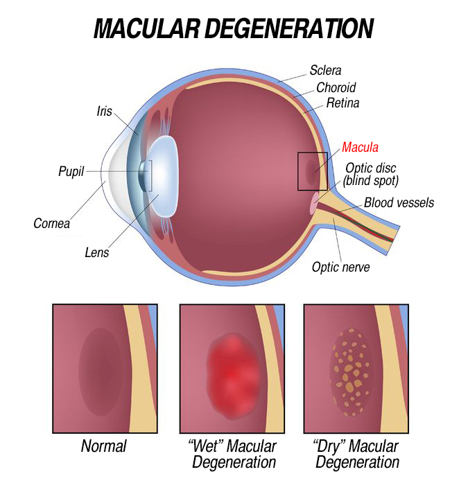
Macular Degeneration is caused by the deterioration of the central portion of the retina, the inside back layer of the eye that records the images we see and sends them via the optic nerve from the eye to the brain. The retina’s central portion, known as the macula, is responsible for focusing central vision in the eye, and it controls our ability to read, drive a car, recognize faces or colors, and see objects in fine detail.

  • Dry macular degeneration (atrophic): When parts of the macula get thinner with age, some small white or yellowish deposits, called drusen, grow on the retina and beneath the macula. You slowly lose central vision.
  • Wet Macular Degeneration (Neovascular): Abnormal blood vessels grow under the retina, they may leak blood or other fluids, and cause scarring of the macula. You lose vision faster than the dry macular degeneration and it is much more serious

2. Symptoms of Macular Degeneration

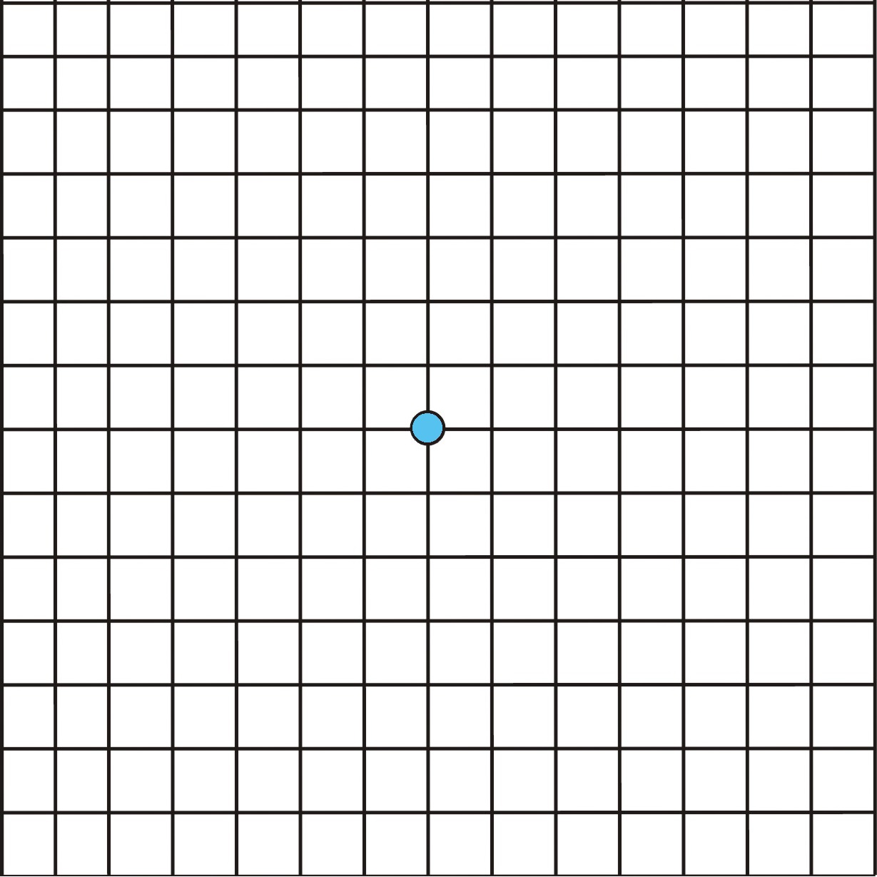
  • Blurry areas appear in the center of your vision
  • A change or distortion in your perception of colour, lines, blind spots
  • A total loss of central visual field

3. How to Prevent Macular Degeneration

  • Don't smoke: Smokers are up to four times more likely than non-smokers to have macular degeneration
  • Obesity control: According to some studies, weight/cholesterol levels may lead to macular degeneration
  • Family history: People with a family history of macular degeneration are at a higher risk
  • Regular eye exams: High Myopia or Aging people

4. Amsler Grid Test for Macular Degeneration

The Amsler grid is used to check whether lines look wavy or distorted, or whether areas of the visual field are missing. It cannot replace a comprehensive eye check up. If you found any distortion or missing lines, please consult your ophthalmologist as soon as possible
  • Tape this page at eye level where light is consistent and without glare. Stand 33cm away from this page
  • If you wear glasses, keep them on
  • Cover one eye and test. Then another eye
  • Look at the blue dot in the center of the grid, try to see if any lines are distorted or missing

5. Treatment Plan for Macular Degeneration

    • Photodynamic Therapy (PDT)
    • Intravitreal Injection

Photodynamic Therapy (PDT): The patient first receives an intravenous injection of a photosensitizer, which is absorbed by the abnormal new blood vessels. After the drugs are absorbed, a laser is used to activate the photosensitizer, thereby destroying the abnormal blood vessels. This is a selective, non-thermal laser treatment that can manage and eliminate choroidal neovascularization in the macular area, helping to control the condition and slow its progression. It is considered a relatively safe therapy. Since age-related macular degeneration can lead to the formation of abnormal blood vessels in the choroid, the photosensitizers used in PDT effectively assist in blocking these abnormal vessels.

Process of Photodynamic Therapy

  1. Fundus Fluorescein Angiography: This examination assesses the condition of the blood vessels in the eye.
  2. Determine the Leakage Situation and Location of the Macular Area: Analyze the leakage in the macular region.
  3. Intravenous Injection of Photosensitizer: Administer the photosensitizer to the patient via intravenous injection, allowing it to be absorbed by the abnormal blood vessels.
  4. Laser Treatment on the Macular Area: Use a laser to irradiate the macular area, activating the photosensitizer to block the abnormal neovascularization beneath the retina.


Precautions:

  • Avoid direct exposure to strong light for 24-48 hours after treatment. It is advisable to wear dark sunglasses to shield your eyes from light.
  • You may experience blurred vision for several hours post-treatment, so do not drive or operate machinery.

Intravitreal Injection: This procedure involves directly injecting anti-VEGF medication into the vitreous of the eye. It inhibits the abnormal growth of blood vessels within the eye, thereby reducing macular edema and bleeding caused by new blood vessels. This helps prevent further bleeding and control the condition. Patients generally need to undergo a series of injections over a specified period, and the treatment shows significant effects; most patients experience improved vision.

Process of Intravitreal Injection:

  1. Eye drops containing anesthetic and antibiotics are applied before the injection.
  2. The doctor will use an eyelid clamp to hold your eye open.
  3. The medication is injected into the vitreous of the eye using a fine needle.

Precautions:

  • Please use antibiotic eye drops or ointment as directed by your doctor.
  • Avoid swimming or engaging in vigorous exercise for one week after treatment.
  • Depending on the treatment plan, you may need to receive another injection of anti-VEGF medication at your scheduled appointment.


Phone
WhatsApp