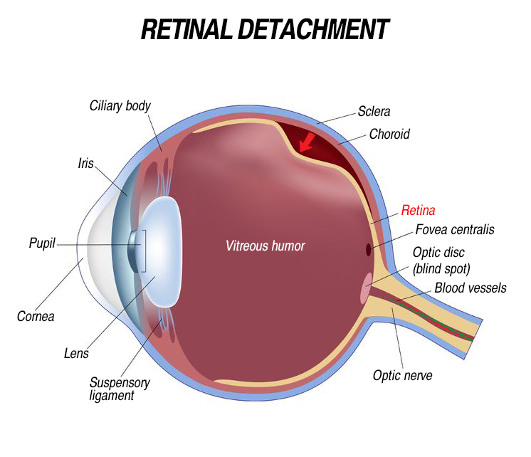
1. What is Retinal Detachment
The retina is a thin and sensitive tissue that is arranged behind the eyeball. Like a film in a camera, the retina converts the optical image into a nerve pulse, transmitted through the optic nerve and seen as the vision in the brain.In most cases, retinal detachment is caused by retinal rupture in the retina, which allows fluid to enter beneath the retinal space, causing the retina to separate from the back of the eyeball. Since the separated retina is partially deprived of blood and nutrient supply, it will degrade and not work properly.
Without prompt treatment, it will lead to Vision damage and blindness in the affected eye.
2. Symptoms of Retinal Detachment
Before this happens, there are signs and symptoms that can alert one to the possible onset of retinal detachment- The sudden appearance of floaters, and they are getting more and more
- Flashes or black shadows
- Partial vision loss
- Sudden onset of blurred or distorted Vision
- If the retina is totally detached, it can cause permanent vision loss
- Some patients have no warning signs at all
3. How to Prevent Retinal Detachment
Retinal detachment can occur at any age, but more common in people over 40 years of age. It usually affects more on men. Retinal detachment is also more likely to occur in the following population:- High myopia (Myopia over -6.00) or Pathological Myopia (Myopia over -10.00)
- Have had a retinal detachment before
- Have a family history of retinal detachment
- Have had cataract surgery
- Have other eye diseases or disorders, such as retinoschisis, uveitis, degenerative myopia, or lattice degeneration
- Have had an eye injury
Pars Plana Vitrectomy (PPV)
- Pars plana vitrectomy (PPV) is a surgical procedure that involves the removal of vitreous gel from the eye. Instruments are introduced into the eye through the pars plana, and injects other fluids (saline liquid, gas or silicone oil) to replace the vitreous humor that ordinarily fills up the inner chambers of the eye. This helps flatten a detached retina and keep it attached to its original position.
Non-PPV Surgery
A gas bubble is injected into the eye, this pushes the wall of the eye against the detached retina, allowing it to re-attach. Then ophthalmologists apply laser or freezing treatment to seal the retinal hole. If there is too much fluid in the bottom area of the retina, ophthalmologists may first remove the fluid and sew one or more silicone bands or tyres to the sclera to enhance the attachment of the retina.
- Pneumatic Retinopexy
- Cryotherapy
- Scleral Encircling & Buckling Surgery